Rib connections
My friend Eric loves a good meal, so to encourage him to read the posts and recognize how awesome ribs are, I'll be starting each post on rib anatomy with an example of ribs from restaurants near the museum!
I told my good friend Eric Scott, a paleontologist who is an expert on fossil horses (and subscriber to this blog), about my plan to write several posts about ribs. He was, shall we say, skeptical. But Eric also loves a good meal, so to encourage him to read the posts and recognize how awesome ribs are, I'll be starting each rib post with an example of ribs from restaurants near the Western Science Center. This week's featured ribs are from the Steer 'n Stein in Hemet. Ribs don't need barbecue sauce to be interesting, but it doesn't hurt!

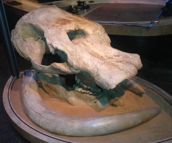
Above is the first right rib of a juvenile Pacific mastodon (Mammut pacificus), seen in posterior view. The bone is still in its plaster field jacket, and has only been prepared on one side. Below is the same bone, with some of the major features labeled:
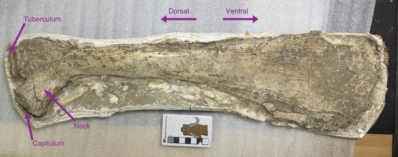
The photo is oriented sideways, so the associated vertebra would have been off the left side of the image. There are two points of articulation with the vertebral column, called the tuberculum and the capitulum. In the more anterior ribs, the capitulum is often offset from the main axis of the rib by a short shaft of bone called the neck. At the other end of the rib, in most mammals and birds and many reptiles the rib will articulate to another rib-like structure called a sternal rib (or, sometimes, "false rib"). The anterior sternal ribs articulate with the sternum, and more posteriorly merge with each other at their tips. However, in the vast majority of mammals the sternal ribs never ossify and remain cartilage through the life of the animal (armadillos and sloths are exceptions that have bony sternal ribs).
As a general rule, the first rib is shorter, straighter, and much broader and flatter in cross-section than the rest of the ribs. This rib is also from a young animal, so the articular surfaces of the tuberculum and capitulum had not yet fused to the rest of the rib (in fact, the capitular articular surface is missing entirely).
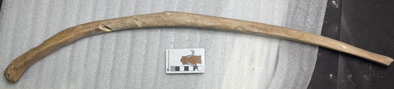
As we move further back in the ribcage, the ribs generally become longer, more curved, and closer to circular in cross-section (although this is extremely variable). The capitulum also tend to become less pronounced, and in the last few sets of ribs is lost entirely, leading to ribs like this mastodon rib:
So these ribs have a tuberculum and in some cases a capitulum that attaches to the vertebral column, but where, exactly? To see that we need to take a look at some thoracic vertebrae with the ribs out of the way. Below are the 2nd and 3rd thoracic vertebrae from a modern white-tailed deer (Odocoileus virginianus):
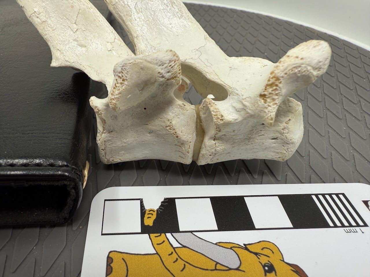
Here's the same image, color coded:
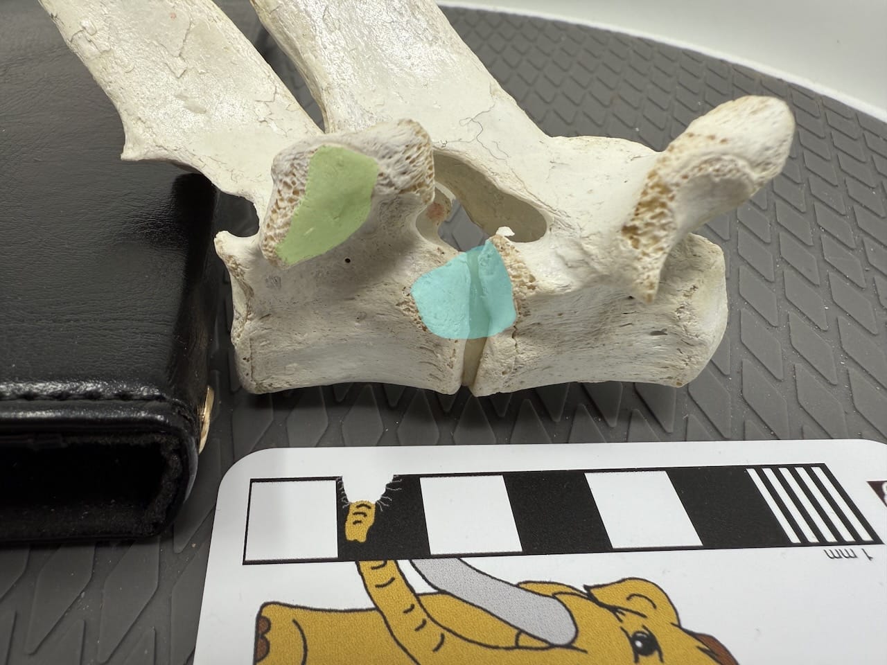
I've highlighted the articular surfaces on the 3rd thoracic, for the 3rd rib. The lime green highlight is the articular facet for the tuberculum, located on the lateral tip of the transverse process facing outward and downward. The cyan highlight is the articulation for the capitulum. This is located low on the front side of the centrum, and the facet actually extends forward onto the back edge of the centrum of the 2nd thoracic. So anterior ribs not only articulate with their corresponding vertebra in two places, but also with the preceding vertebra in one place! Here's what this looks like with the rib in place:
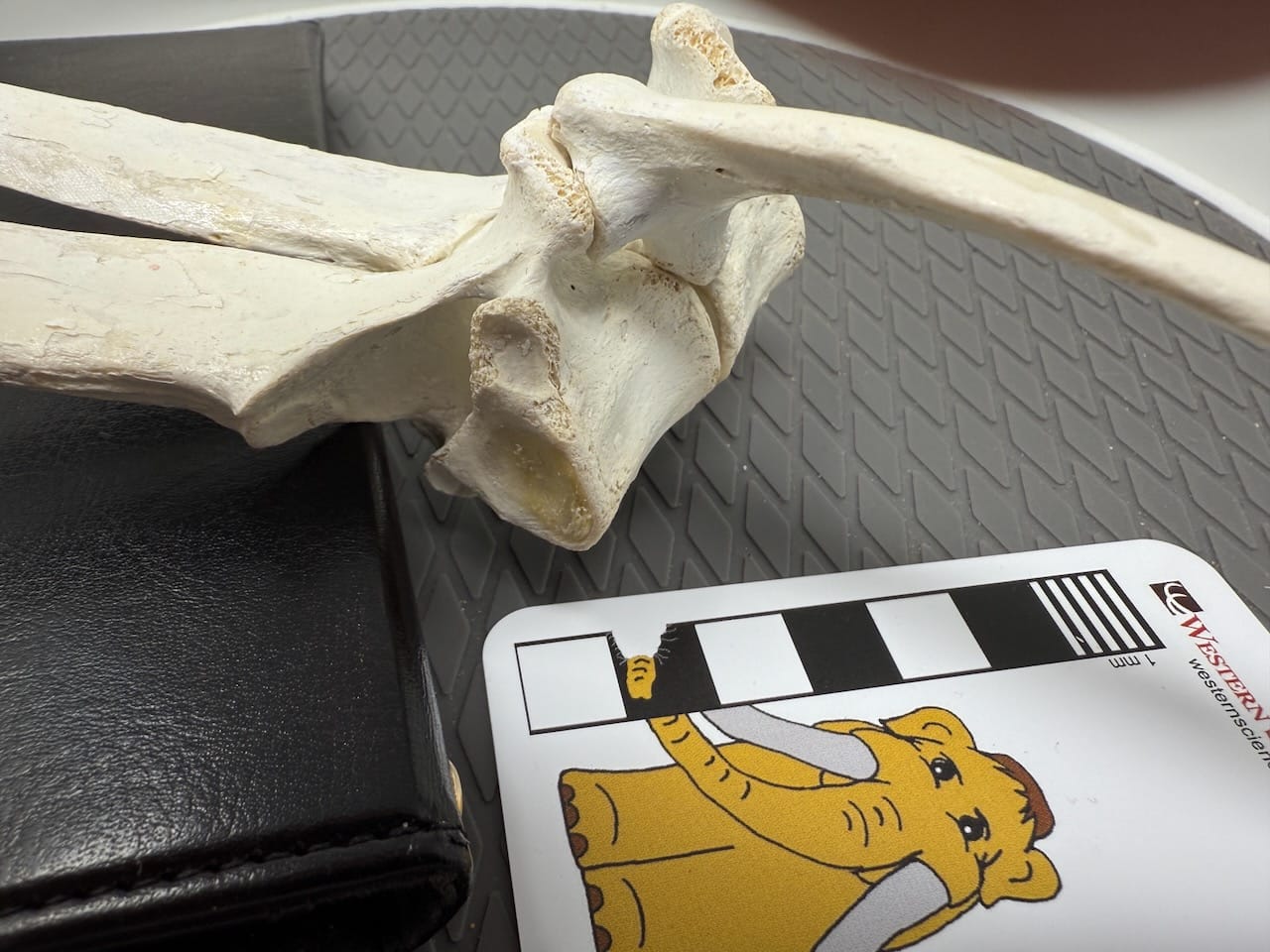
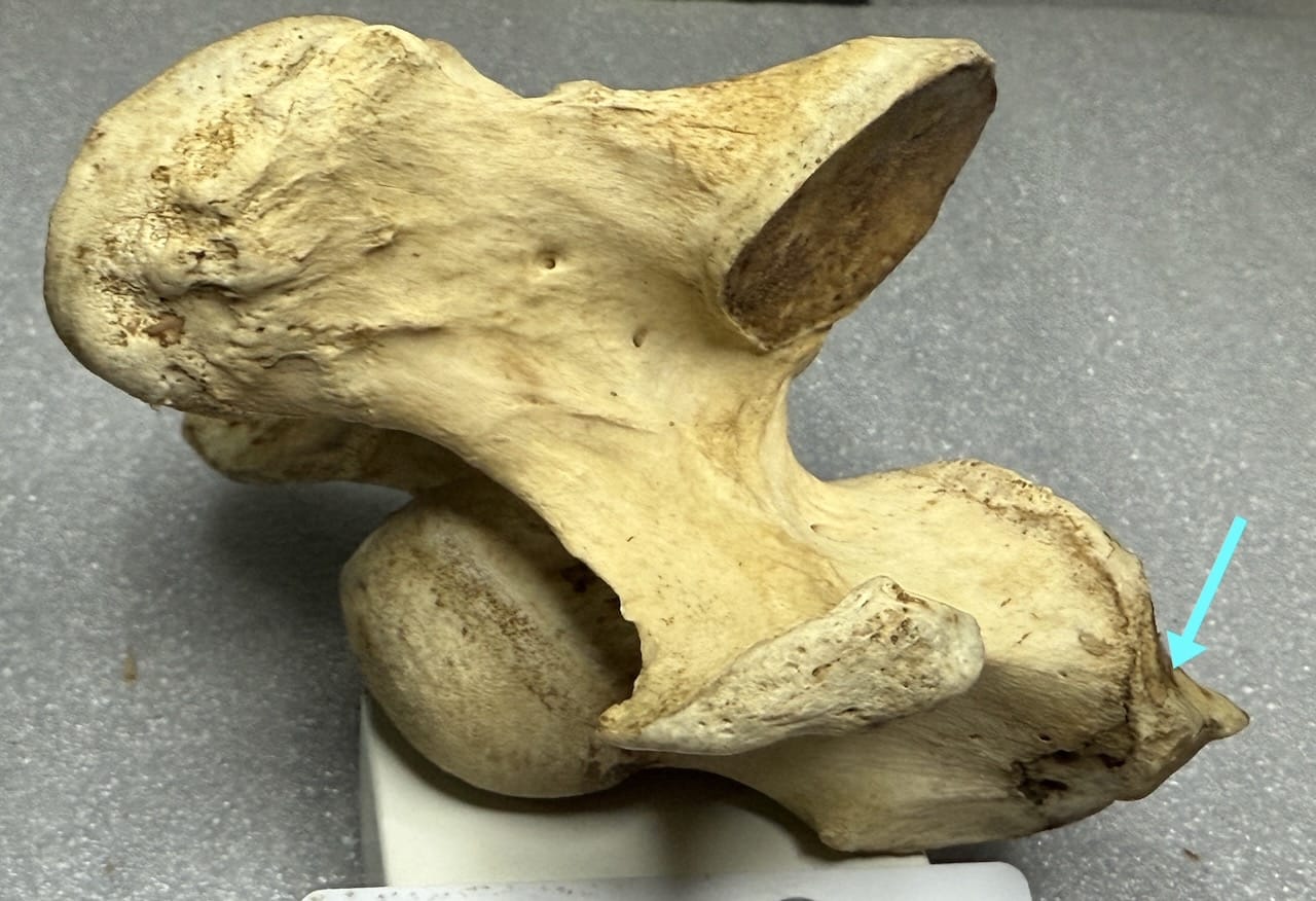
Posteriorly, the capitulum is lost, and the articular facets on the vertebral centra are lost as well; these vertebrae only have the articulations for the tuberculum at the ends of the transverse processes. In mammals, which lack ribs on the neck (cervical) vertebrae, the capitular articulation also leads to an unusual situation in which the last (7th) cervical vertebra has capitular facets on the posterior edge of the centrum, but because it has no corresponding rib there are no facets on the anterior edge of the centrum, nor are there tubercular facets on the transverse processes. The 7th cervical is the only vertebra with this pattern, as seen in the example below from a horse:
The ribs serve all kinds of functions, including protecting the internal organs, providing support for the forelimbs, and a host of other things, sometimes with unique functions in different groups (we'll look at some examples starting next week). But a major function in mammals and most reptiles, that depends on the ribs' shape and articular style, is breathing.
There are a series of muscles called intercostal muscles that connect the ribs to each other (the yummy parts in the picture at the top). When the intercostal muscles contract, they tend to pull the ribs forward. But because the ribs articulate at their top ends, and because they're curved, as they pull forward they also rotate outward, increasing the volume of the thoracic cavity where the lungs are located. This lowers the pressure in the lungs, pulling in air from outside. This is called "costal respiration" and is responsible for shallow breathing in mammals; it's the primary means of breathing in some reptiles. This is why it hurts so much to breathe if you break ribs; they're moving back and forth all the time, with every breath. Our ability to live is largely dependent on the shape and articular style of our ribs!
We'll continue with ribs next week, with some specific examples of how ribs have been modified in some animals to serve additional functions. And, if you're near Hemet, try out the ribs at Steer 'n Stein!

If you like what you're reading, please consider becoming a paid subscriber or leaving a tip. All proceeds go to cover the cost of maintaining the site and supporting research and education at the Western Science Center.