The Tuna's Tail
Before coming to the Western Science Center I worked at the Virginia Museum of Natural History as Curator of Paleontology. My primary field site during that time was the Carmel Church Quarry in eastern Virginia, a remarkable location with multiple fossiliferous beds spanning 60 million years of time. During my time in Virginia I ran approximately 50 excavations at Carmel Church, recovering many thousands of fossils. By far the most remarkable unit there was the middle Miocene (14-million-year-old) Calvert Formation, which produced at least 80 different species of animals dominated by whales, sharks, and bony fish.
When I moved to California and became an administrator, much of my research had to take a backseat. But recently I've been able to pick up the threads of some of the unfinished projects, and we recently sent a WSC team to Virginia to borrow some of the specimens from my old excavations. These included some of the Carmel Church fish.
Other than the whales, most Carmel Church remains are isolated bones. So I was quite excited when, on 5 April 2010, we found what appeared to be six associated fish vertebrae!
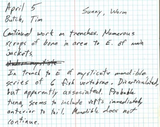
I took a photo as we uncovered them:
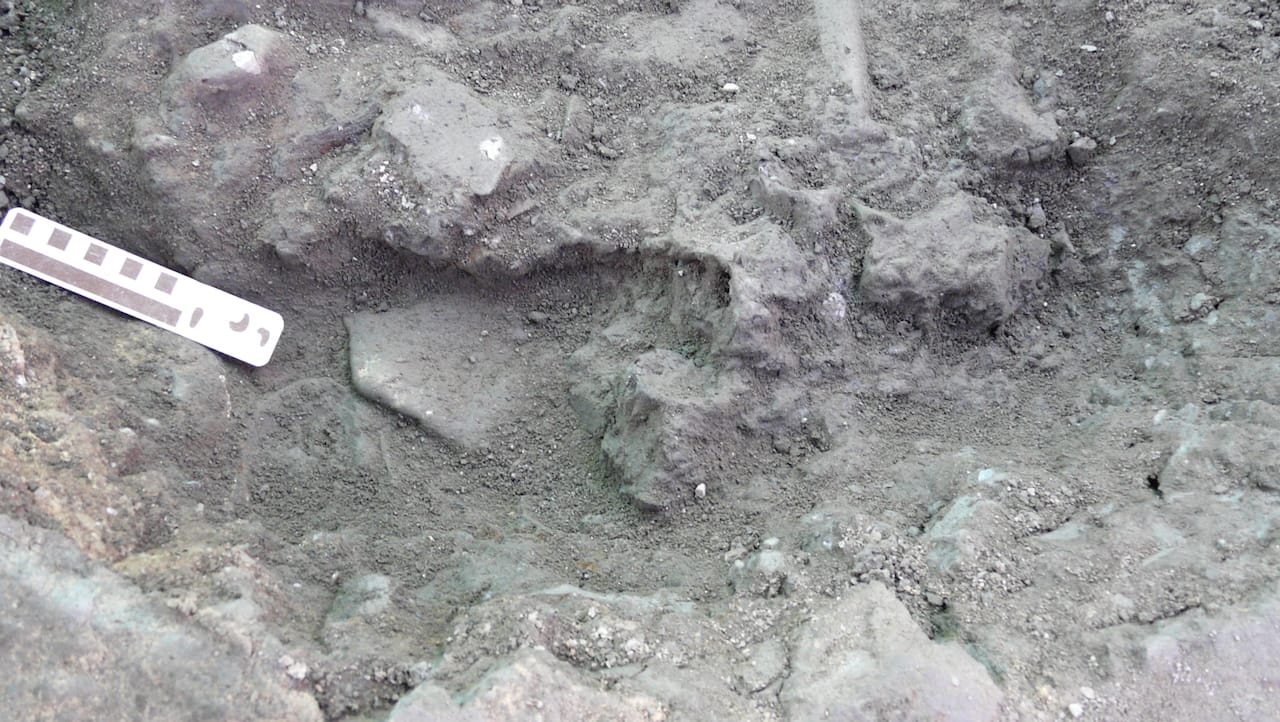
And here's the same image with the six fish vertebrae circled in red (almost all the other flat-ish surfaces are whale bones):
After getting these back to the museum and cleaning them, it turned out there were 10 vertebrae instead of 6, and they formed a continuous series:
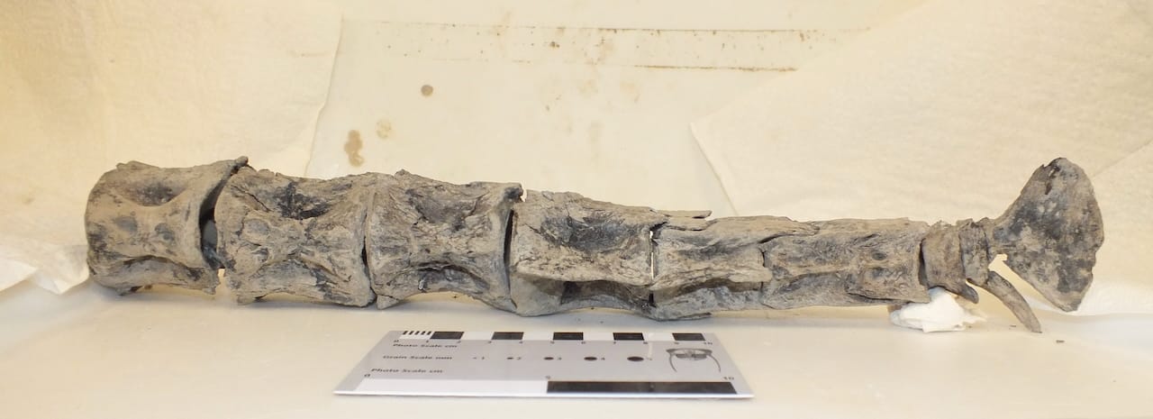
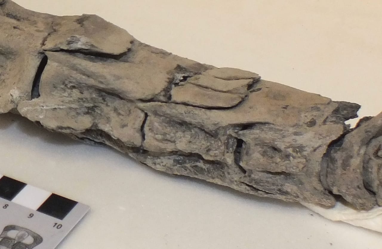
The neural spines and haemal spines are missing on the first 3 vertebrae, but they are present on the next four. In these four they are highly modified, angled straight back and locking into grooves on the centrum of the next vertebra back. This makes a rigid, interlocking box structure, that is so tight that these vertebrae couldn't be separated even after cleaning them, as is visible in this closeup of the underside:
This particular interlocking box structure is typical of the "true tunas" in the genus Thunnus, so it turned out my tentative field identification in the notebook was correct. The triangular terminal vertebra, called the hypural (actually formed by the fusion of about 6 separate bones) also has a shape typical of Thunnus; we had already recovered several tuna hypurals at Carmel Church.
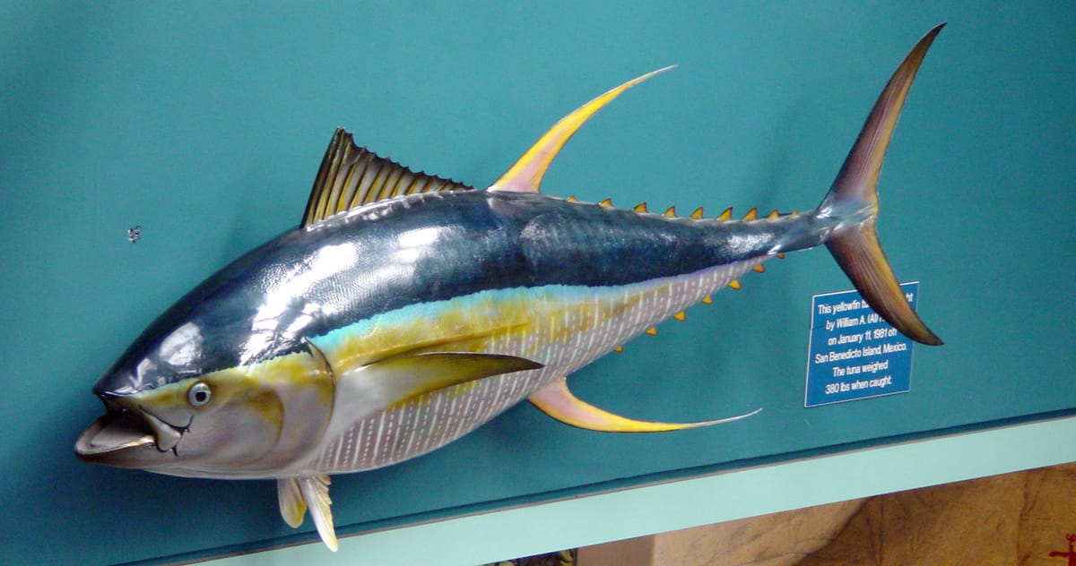
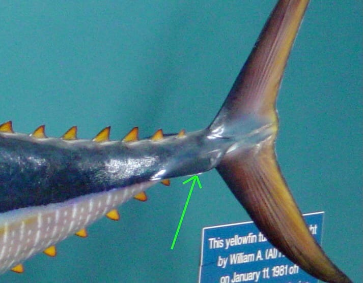
Another fun feature of tuna are the strong lateral keels at the base of the tail, visible on the the taxidermy specimen at the top of the post (closeup below, green arrow indicated the keel):
These keels are actually supported by lateral processes on the vertebrae in the box structure, visible when seen from above (starting around the 3.5 cm mark on the scale bar):
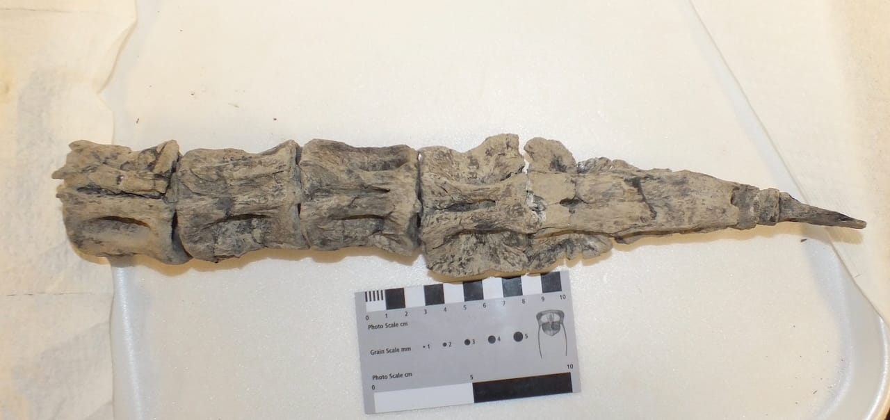
So how much of this tuna did we actually recover? Thunnus seems to pretty consistently have 39 vertebrae, and we have the last 10. So we recovered the green-outlined section on the skeleton shown below:
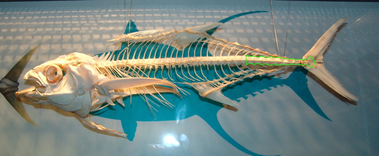
Based on the size of our vertebrae, that translates to a tuna roughly 2.5 m in length. That's respectably large for a tuna, but not gigantic; some modern tuna species can reach up to around 4.5 m. On the other hand, we recovered individual vertebrae at Carmel Church that were at least twice the diameter of the largest vertebra in this tail series, suggesting the Carmel Church was home to tuna that approached the size of the largest living tuna.
At Western Science Center we've digitized this tuna tail, and a 3d print will be included in an upcoming exhibit in our Mobile Museum.

If you like what you're reading, please consider becoming a paid subscriber or leaving a tip. All proceeds go to cover the cost of maintaining the site and supporting research and education at the Western Science Center.