Tracing holes in bones
When trying to understand the anatomy of a bone, one of the first steps is identifying structures that are shared with the same bone from other species (the technical term is "homologous structures"). One that is quite useful to look for is holes in the bone, called foramina (singular: foramen) that allow nerves and blood vessels to pass through the bone. Of course, the skull has lots of nerves and blood vessels, so it also have a number of interesting foramina, including the infraorbital foramen.
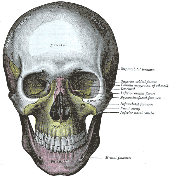
As you can see from the drawing of a human skull above, the infraorbital foramen is located in the maxilla, the bone that holds most of our upper teeth and forms most of our lower face. The infraorbital foramen leads to a canal that opens into the bottom of our eye socket (or orbit, hence the name). The infraorbital nerve, infraorbital artery, and infraorbital vein all pass through the foramen and then split into numerous small branches that spread out across the maxilla, supplying blood and sensory input to the tissues on our lower face. The infraorbital nerve is responsible for our senses on our lower eyelid, the side of our nose, our upper front teeth, and our upper lips.
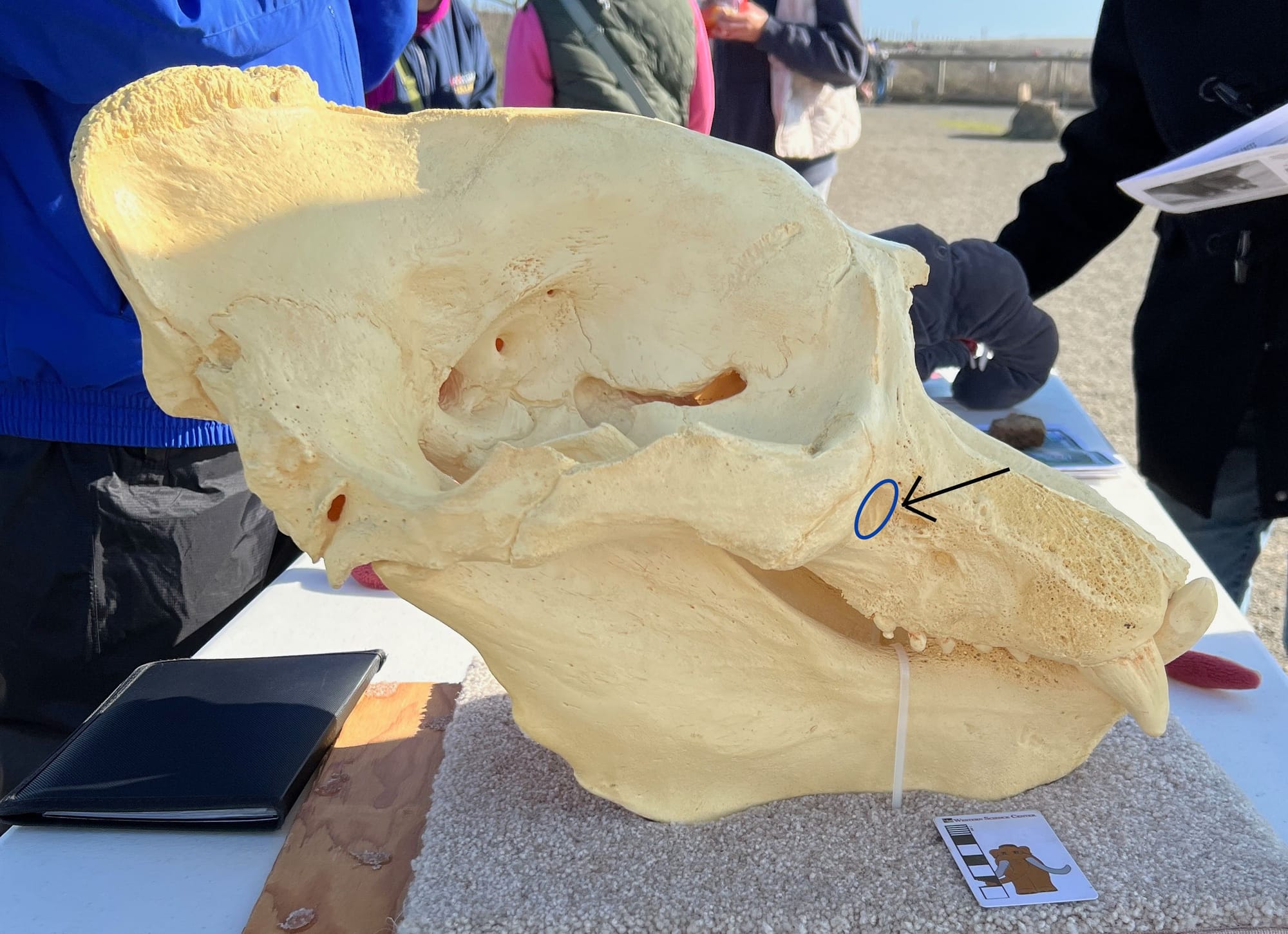
Nearly all mammals have a maxillary foramen, but its expression can vary greatly. In humans it is pretty small, in part because our faces are short without a lot of soft tissues, and in spite of the sensitivity of our lips, compared to many animals we don't have a lot of sensory apparatus located on our faces. But many animals have a lot of sensory organs on their faces, such as the whiskers on this elephant seal:
Sleeping male elephant seal
These numerous, sensitive whiskers need a large nerve and a good blood supply, so there is a correspondingly large infraorbital foramen, indicated by the circle and arrow:
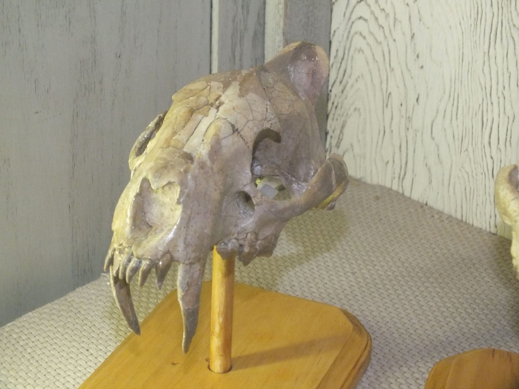
Like the elephant seals, most carnivorans have very large infraorbital foramina, such as the sabertooth nimravid Hoplophoneus:
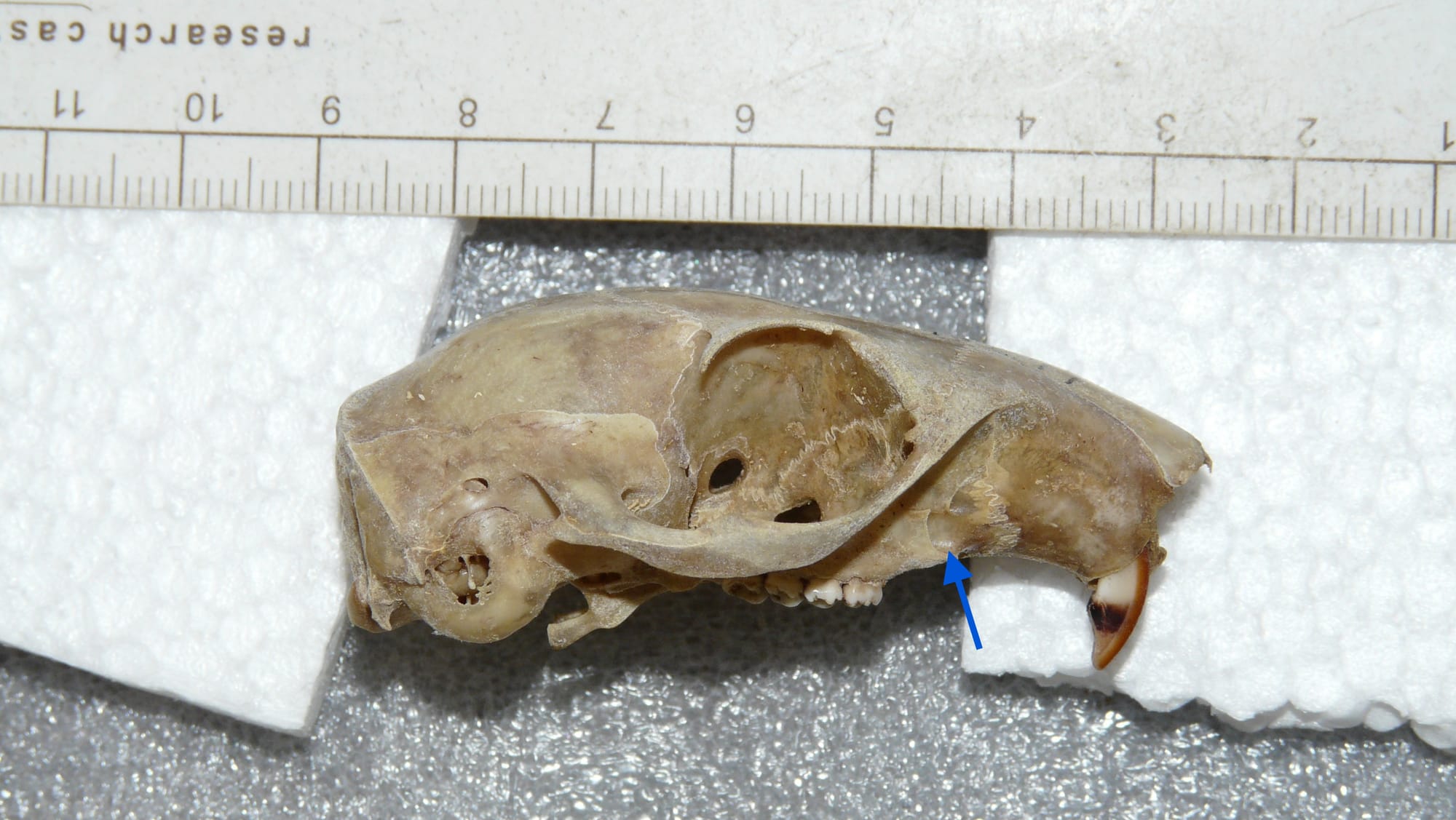
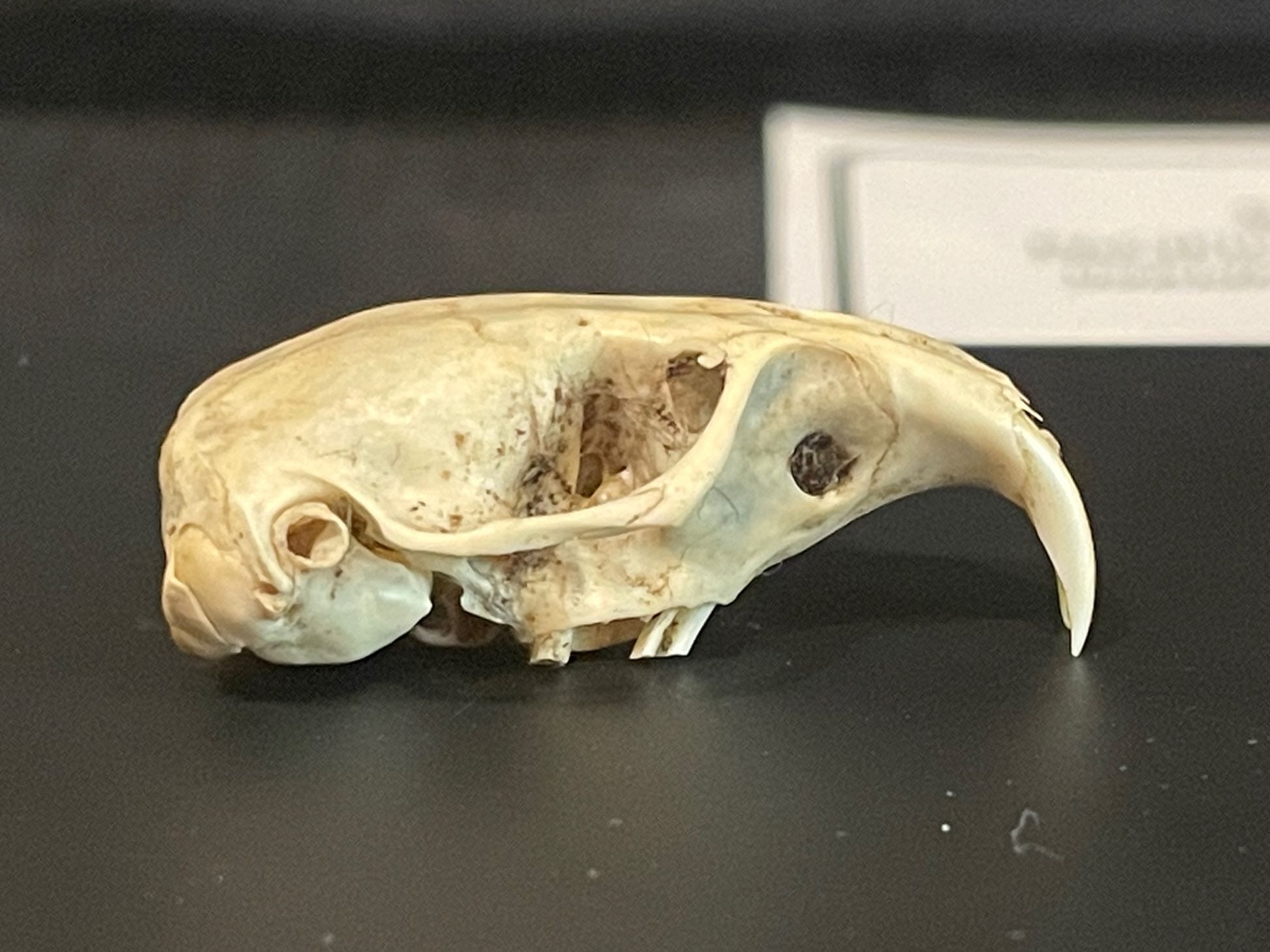
Most rodents, such as this Eastern fox squirrel (Sciurus niger), have a moderately large, slot-like infraorbital foramen that opens anteriorly:
However, one rodent group, the geomyoids, have a very large infraorbital canal that opens the the side instead of anteriorly, as the the pocket gopher (Thomomys bottae) below. This is one of the major identification features of the geomyoids.
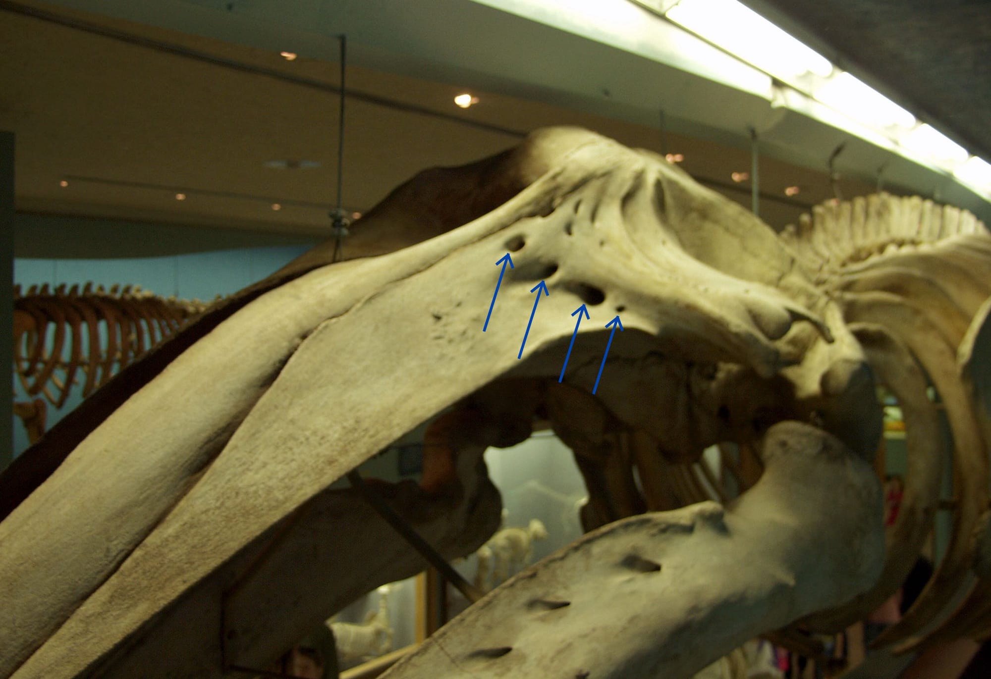
Baleen whales have extensive upper jaws that make up about 80% of their skulls. The infraorbital canal actually splits into multiple branches inside the maxilla and comes out as several different foramina. However, as baleen whales have no upper teeth, and very little in the way of sensory structures on the face, the foramina are very small compared to the size of the skull.
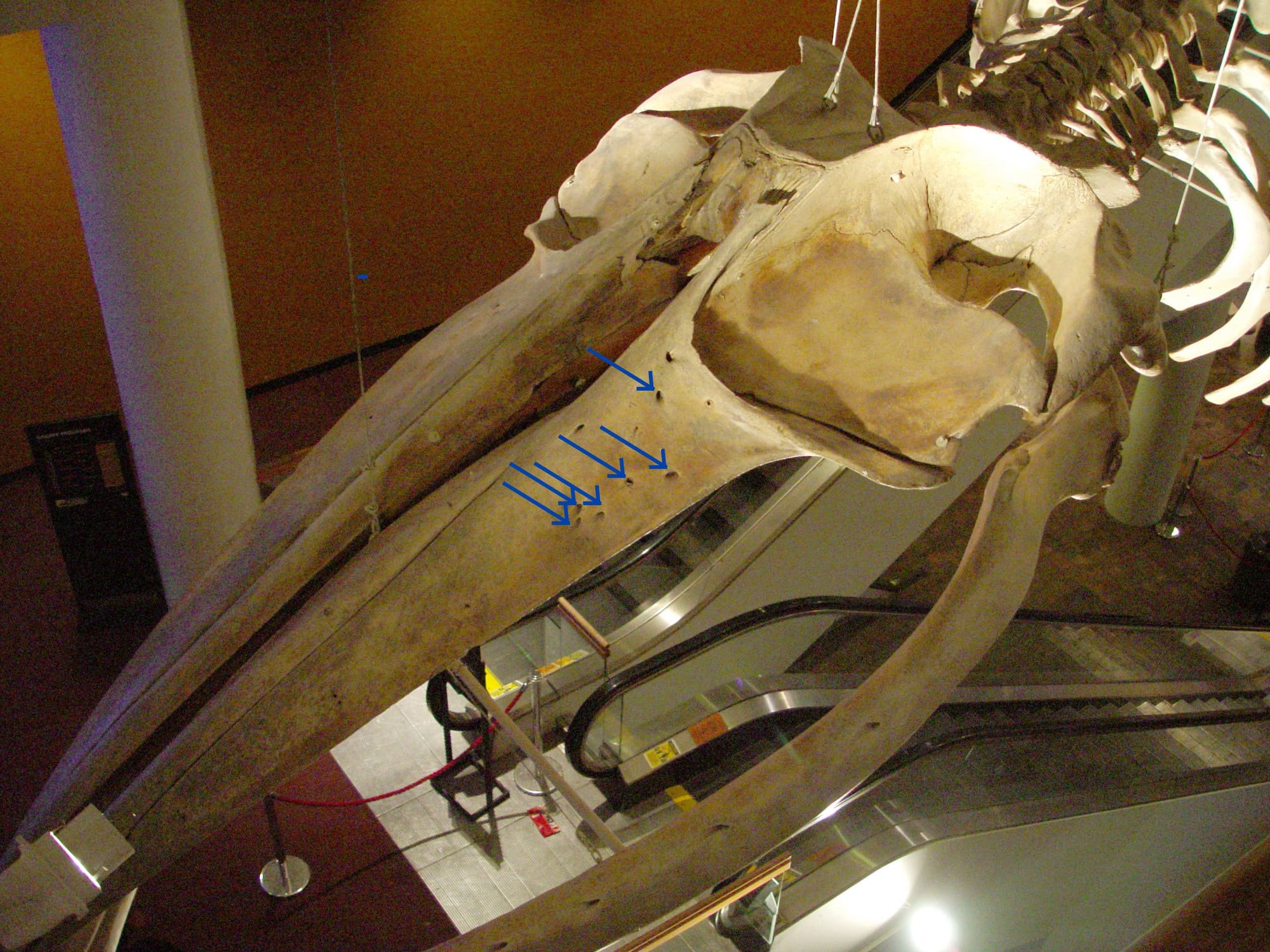
But even in baleen whales there's variation. The toothed ancestors of baleen whales had relatively much larger infraorbital foramina, and the modern gray whale (Eschrichtius robustus), which does have a few sensory whiskers on its snout, has very large infraorbital foramina.
Small details in bones such as the size and position of foramina can tell us a great deal about the biology and relationships of vertebrates, which is why zoologists and paleontologists put so much effort into documenting them.

If you like what you're reading, please consider becoming a paid subscriber or leaving a tip. All proceeds go to cover the cost of maintaining the site and supporting research and education at the Western Science Center.

